Corneal Topography – Painless Imaging!
What is a Cornea!
The cornea is the outermost lens of the eye. It works like a window, controlling and focusing the amount of light that enters the eye. The cornea is responsible for 65-75 percent of the entire focusing capacity of the eye. Light rays must be focused by the cornea and lens to fall precisely on the retina for you to see clearly.
The Normal Cornea
An eye with normal vision has a uniformly rounded cornea, but vision is “less-than-perfect” if the cornea is excessively flat, too steep, or unevenly curved. In situations like these, we must assess the cornea to treat diseases or abnormalities caused by corneal malfunctions.
Why Corneal Imaging!
But we are unable to photograph a corneal function, even though our goal is to learn how the cornea of a specific patient is functioning. What we can do, however, is analyze the structural intricacies of the cornea in detail such that we can infer indirectly from our obtained information how a particular cornea is working or will work in the future.
Corneal imaging works on this principle. Without a doubt, the more extensive the structural study, the more precise our diagnosis and, as a result, treatment will be. This necessitates data gathering and analysis.
The application of digital analysis with superfast computers has served us a long way to achieve our goal which was not possible until a few decades earlier.
What is Corneal Topography?
Corneal topography is a computer-assisted diagnostic test for generating a three-dimensional map of the cornea's surface curvature. About 70% of the focusing power of the eye comes from the cornea (the front window of the eye). An eye with normal vision has a uniformly rounded cornea, but vision is “less-than-perfect” if the cornea is excessively flat, too steep, or unevenly curved. The most significant benefit of corneal topography is its ability to detect abnormal conditions that are undetectable by most other conventional tests.
Corneal topography produces a detailed visual description of the cornea's shape and power. This type of examination gives your doctor extremely minute details about the condition of your corneal surface. These details are utilized to diagnose, monitor, and treat a variety of eye problems. They're also utilized for contact lens fitting and surgery planning, including laser vision correction. The corneal topography map is used in conjunction with other tests to establish how much corneal tissue will be removed to correct vision and with what ablation pattern for laser vision correction.
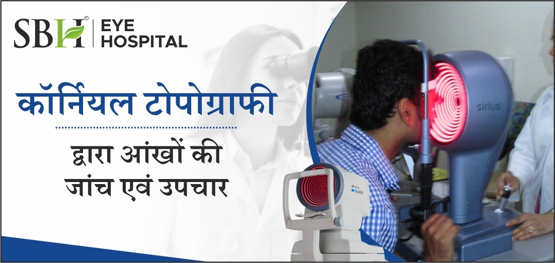
How is a Corneal Topography Performed?
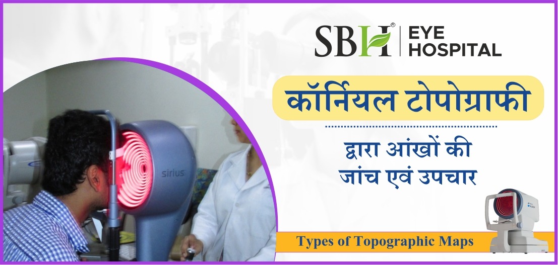
A corneal topography test is a quick and painless procedure. You will sit in front of an illuminated bowl with a pattern of rings and rest your head against a bar during the test. On a computer screen, a series of data points will be collected, and a color-coded image of your corneal shape will be generated.
Types of corneal topography
Corneal Topography is performed using three different technologies:
Placido Disc Topography
The curvature, irregularity, tear film quality, foreign substances, and other parts of the anterior cornea are measured using the Placido disc reflection systems. The reflection is highly dependent on the tear film reflecting the light, which can be either small-cone or large-cone. Small cones are more accurate since they gather more data points, but huge cones are more user-friendly and make data collection much easier.
Scheimpflug and Scanning-slit Topography
These two systems provide information about the anterior and posterior cornea and, are used to identify, treat, and manage corneal swelling, which is particularly critical for contact lens wearers.